Primary Lymphoid Organs
Immature lymphocytes generated in hematopoiesis mature and become committed to a particular antigenic specificity within the primary lymphoid organs. Only after a lymphocyte has matured within a primary lymphoid organ is the cell immunocompetent(capable of mounting an immune response). T cells arise in the thymus,and in many mammals – humans and mice for example – B cells originate in bone marrow.
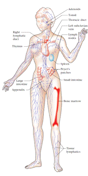
Figure 1. The human lymphoid system. The primary organs (bone marrow and thymus) are shown in red; secondary organs and tissues, in blue. These structurally and functionally diverse lymphoid organs and tissues are interconnected by the blood vessels (not shown) and lymphatic vessels (purple) through which lymphocytes circulate. Only one bone is shown, but all major bones contain marrow and thus are part of the lymphoid system. [Adapted from H. Lodish et al., 1995, Molecular Cell Biology, 3rd ed., Scientific American Books.]
THYMUS
The thymus is the site of T-cell development and maturation. It is a flat, bilobed organ situated above the heart. Each lobe is surrounded by a capsule and is divided into lobules, which are separated from each other by strands of connective tissue called trabeculae. Each lobule is organized into two compartments: the outer compartment, or cortex, is densely packed with immature T cells, called thymocytes, whereas the inner compartment, or medulla, is sparsely populated with thymocytes.
Both the cortex and medulla of the thymus are crisscrossed by a three-dimensional stromal-cell network composed of epithelial cells, dendritic cells, and macrophages, which make up the framework of the organ and contribute to the growth and maturation of thymocytes. Many of these stromal cells interact physically with the developing thymocytes (Figure 2). Some thymic epithelial cells in the outer cortex, called nurse cells,have long membrane extensions that surround as many as 50 thymocytes, forming large multicellular complexes. Other cortical epithelial cells have long interconnecting cytoplasmic extensions that form a network and have been shown to interact with numerous thymocytes as they traverse the cortex.
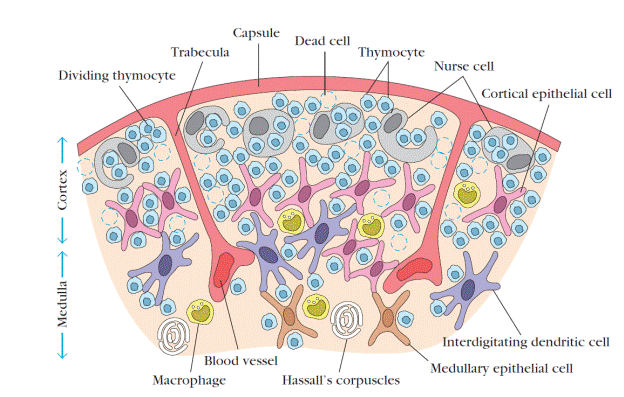
Figure 2. Diagrammatic cross section of a portion of the thymus, showing several lobules separated by connective tissue strands (trabeculae). The densely populated outer cortex is thought to contain many immature thymocytes (blue), which undergo rapid proliferation coupled with an enormous rate of cell death. Also present in the outer cortex are thymic nurse cells (gray), which are specialized epithelial cells with long membrane extensions that surround as many as 50 thymocytes. The medulla is sparsely populated and is thought to contain thymocytes that are more mature. During their stay within the thymus, thymocytes interact with various stromal cells, including cortical epithelial cells (light red), medullary epithelial cells (tan), interdigitating dendritic cells (purple), and macrophages (yellow). These cells produce thymic hormones and express high levels of class I and class II MHC molecules. Hassalls corpuscles, found in the medulla, contain concentric layers of degenerating epithelial cells. [Adapted, with permission, from W. van Ewijk, 1991, Annu. Rev. Immunol. 9:591, © 1991 by Annual Reviews.]
The function of the thymus is to generate and select a repertoire of T cells that will protect the body from infection. As thymocytes develop, an enormous diversity of T-cell receptors is generated by a random process that produces some T cells with receptors capable of recognizing antigen-MHC complexes. However, most of the T-cell receptors produced by this random process are incapable of recognizing antigen-MHC complexes and a small portion react with combinations of self antigen-MHC complexes. The thymus induces the death of those T cells that cannot recognize antigen-MHC complexes and those that react with self-antigen-MHC and pose a danger of causing autoimmune disease. More than 95% of all thymocytes die by apoptosis in the thymus without ever reaching maturity.
Дата добавления: 2016-07-18; просмотров: 2115;
Поиск по сайту
Узнать еще
- CANCERS OF DEFINITE ORGANS
- Cutaneous-Associated Lymphoid Tissue
- MUCOSAL-ASSOCIATED LYMPHOID TISSUE
- Primary (essential) hypertension
- Primary Lymphoid Organs
- Secondary Lymphoid Organs
- Таким образом, чтобы обратится блочному устройству на мастер диску к первой primary таблице нужно обратится к специальному файлу в директории /dev

Публикации по технике и механике

Публикации по биологии

Публикации по информатике

Публикации по строительству

Публикации по физике

Публикации по химии

Публикации по электронике

Публикации по искусству

Публикации по географии

Публикации по медицине
