THE FIRST WEEK of LIFE. FERTILIZATION to IMPLANTATION PERIOD
Cleavage.Cleavage of the zygote occurs as the fertilized ovum moves passively toward the uterus. The cleavage type is:
· the holoblastic (complete),
· unequal(blastomeres are different in size),
· asynchronous (blastomeres are divided at different time)
The first cleavage division is meridian with the respect to the animal-vegetative axis and results in two blastomeres of unequal size. The larger cell divides next, giving a three-cell stage. Thereafter the divisions are irregular with the formation of a mulberry-shaped mass of blastomeres - the morula of 16-32 cells (Fig.24). Some of the cells are smaller and light (they'll give rise to trophoblast), another cells are larger and dark (they give rise to embryoblast). The morula is surrounded by the zona pellucida. This occurs during the 3-4 days taken for passage through the uterine tube.
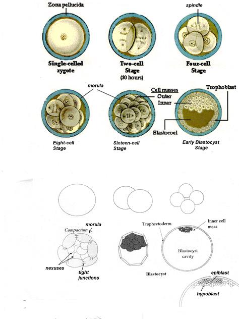 Fig.24.
During next 3-4 days, while free in the uterine cavity, the morula develops into a blastocyst. The cells of the morula are highly active and undergo the changes of shape and relative position. While still within the zona pellucida, fluid-filled spaces appear between the centrally placed cells of the morula; these space soon coalesce to form a blastocystic cavity. The origin of the fluid has been ascribed to endosmosis of uterine fluid, to secretion by the blastomeres, or to central cellular degeneration. The resulting blastocyst has an external wall of primary trophoblast within which is an eccentrically-place inner cell mass, or embryoblast, bulged into the cavity. The primary trophoblast is the first extra-embryonic membrane. Cells of the trophoblast are connected by the tight junctions, within the embryoblast cells are interact by gap junctions (nexuses). Certain formative cells of the embryoblast will form the body of the embryo while all remaining blastocystic cells will form extra-embryonic membranes. The area of contact between the embryoblast and the overlying polar trophoblast defines the embryonic pole of the blastocyst. The blastocyst remains in the lumen of the uterus for 2 or 3days and comes into contact with the surface of the endometrium, immersed in the secretion of the endometrial glands. The day 5, preimplantation human embryo contains 200 to 250 cells, only 30 to 34 of which are inner cell mass cells.
On the 6 or 7 day after fertilization the zona pellucida disappears, allowing cells of the trophoblast, which have the capacity to invade the mucosa, to come into direct contact with the endometrium. Immediately thereafter, the cells of the trophoblast begin to multiply, ensuring, with the help of the endometrium, the nourishment of the embryo.
Implantation (Fig.25) involves penetration through the uterine epithelium.
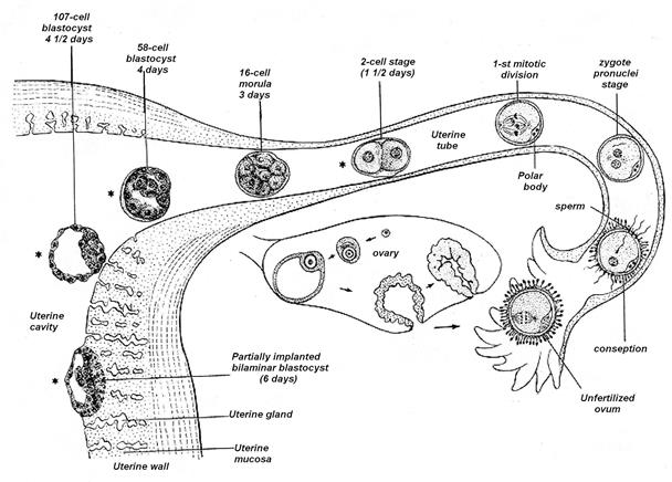
Fig.25
Implantation steps are (Fig.26):
· Apposition: of blastocyst to endometrial epithelium; attachment occurs at the embryonic pole, probably around 6 days; attachment usually occurs between the surface openings of the uterine glands.
· Adherence: via cell adhesion molecules
· Formation of Syncytiotrophoblast: Fusion of cytotrophoblast cells results in giant multinucleated symplast that will surround complete embryo
· Penetration: syncytiotrophoblast is invasive and works way into uterine tissue ultimately making contact with maternal blood vessels
· Decidual Reaction: uterine tissue responds to embryonic invasion by setting up an immunological barrier, the decidua (because the fetus has a different genetic makeup than the mother). The stromal cells enlarge and become pale as glycogen and lipid droplets collect in the cytoplasm.
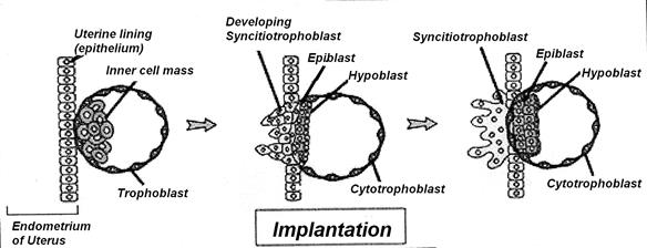
Fig.26.
Дата добавления: 2020-05-20; просмотров: 967;
Поиск по сайту
Узнать еще

Публикации по технике и механике

Публикации по биологии

Публикации по информатике

Публикации по строительству

Публикации по физике

Публикации по химии

Публикации по электронике

Публикации по искусству

Публикации по географии

Публикации по медицине
