THE SECOND WEEK of LIFE. BILAMINAR PERIOD.
This type of interstitial implantation occurs in humans and a few other mammals. The process starts around the7-th day, and on about the 9-th day the embryo is totally submerged in the endometrium, from which it will receive protection and nourishment during the pregnancy. Within implantation cavity, the blastocyst is in fluid medium consisting of extravasated blood, uterine milk and the cytolytic products which follow the breakdown of surface and glandular epithelia, decidual cells and vascular endothelium. The syncytiotrophoblast engulfs this material - thus, histiotrophic type of the embryo' nutrition occurs during the first 2 weeks.
During implantation, the trophoblast differentiates into 2 layers: the syncytiotrophoblast (or, symplastotrophoblast) and the cytotrophoblast (Fig.26). The cytotrophoblastconsists of an irregular layer of mononucleated ovoid cells. The syncytiotrophoblastis a multinucleated external layer (symplast) arises from the fusion of mononucleated cytotrophoblast cells. So, the cytotrophoblast expands mitotically into the syncytiotrophoblast to form primary chorionic villi(composed of trophoblast only). [Cells from these villi can be removed for early genetic testing at some risk to the fetus (chorionic villus sampling)]
The syncytiotrophoblast superficial cytoplasm contains vesicles covered by smooth membranes. The syncytiotrophoblast has active invasive properties and erodes maternal epithelia, stroma and blood vessels to form extra-cytoplasmic cavities. These cavities increase in size and communicate with one another, resulting in a spongy structure. Thus, lacunae are formed, lined with syncytiotrophoblast. The lytic activity of the syncytiotrophoblast causes the rupture of both arterial and venous maternal blood vessels, with overflow of blood into these lacunar spaces. Blood flows from the arterial vessels to the lacunae and from there to the veins. As the conceptus enlarges, histiotrophic nutrition diminishes and haemotrophic nutrition begins.
Gastrulation. In mammals gastrulation occurs by ways of combination of delaminationandimmigration. The first phase of gastrulation – the delamination coincides in time with the process of implantation and results in formation of the bilaminar disk-shaped mass of the cells – germ disk:
· the upper layer of the disk called the epiblast consists of high columnar cells;
· the lower layer called the hypoblast consists of cuboidal cells.
• The epiblast subsequently gives rise to all 3 germ layers of the embryo.
• The hypoblast does not take part in the formation of the embryo body proper and is later displaced to extra-embryonic regions
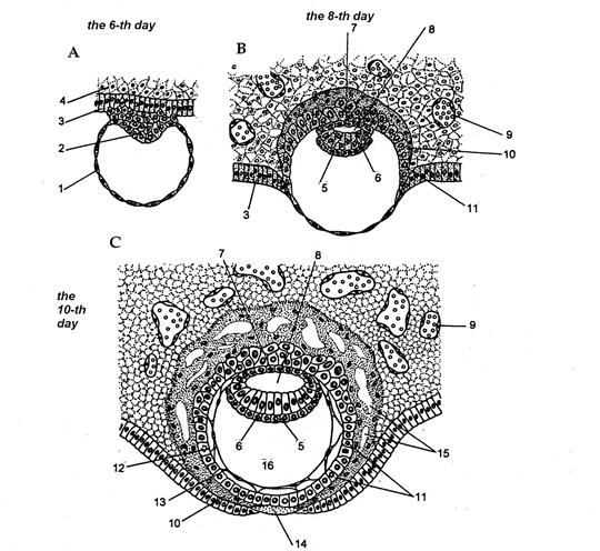
Fig.27.
As implantation proceeds, Epiblast cells cavitate to form the amniotic vesicle; Hypoblast cells migrate and will form the yolk sac. In humans the yolk sac contains no yolk but is important for the transfer of nutrients between the fetus and mother. The cells forming the amniotic wall are called the amnioblasts or amniotic epithelium. The wall of the yolk sac is called the yolk extra-embryonic endoderm. The epiblast forms the floor of the amniotic cavity, whereas the hypoblast represents the roof of the yolk sac.
The loosely arranged cells called the extra-embryonic mesoderm (mesoblast) differentiate and surround the amnion and the yolk sac stabilizing their wall. Extraembryonic somatic mesoderm lines the cytotrophoblast and covers the amnion. Extraembryonic somatic mesoderm also forms the connecting stalk. Extraembryonic visceral mesoderm covers the yolk sac.
When the extraembryonic mesoderm grows into the primary villi, they become the secondary chorionic villi(Fig.28 ).

Дата добавления: 2020-05-20; просмотров: 899;
Поиск по сайту
Узнать еще

Публикации по технике и механике

Публикации по биологии

Публикации по информатике

Публикации по строительству

Публикации по физике

Публикации по химии

Публикации по электронике

Публикации по искусству

Публикации по географии

Публикации по медицине
