THE THYMUS AND IMMUNE FUNCTION
The role of the thymus in immune function can be studied in mice by examining the effects of neonatal thymectomy, a procedure in which the thymus is surgically removed from newborn mice. These thymectomized mice show a dramatic decrease in circulating lymphocytes of the T-cell lineage and an absence of cell-mediated immunity. Other evidence of the importance of the thymus comes from studies of a congenital birth defect in humans (DiGeorge’s syndrome)and in certain mice (nude mice)in which the thymus fails to develop. In both cases, there is an absence of circulating T cells and of cell-mediated immunity and an increase in infectious disease.
Aging is accompanied by a decline in thymic function. This decline may play some role in the decline in immune function during aging in humans and mice. The thymus reaches its maximal size at puberty and then atrophies, with a significant decrease in both cortical and medullary cells and an increase in the total fat content of the organ. Whereas the average weight of the thymus is 70 g in infants, its age-dependent involution leaves an organ with an average weight of only 3 g in the elderly (Figure 3).
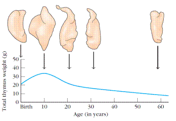
Figure 3. Changes in the thymus with age. The thymus decreases in size and cellularity after puberty.
A number of experiments have been designed to look at the effect of age on the immune function of the thymus. In one experiment, the thymus from a 1-day-old or 33-monthold mouse was grafted into thymectomized adults. (For most laboratory mice, 33 months is very old.) Mice receiving the newborn thymus graft showed a significantly larger improvement in immune function than mice receiving the 33-month-old thymus.
BONE MARROW
In humans and mice, bone marrow is the site of B-cell origin and development. Arising from lymphoid progenitors, immature B cells proliferate and differentiate within the bone marrow, and stromal cells within the bone marrow interact directly with the B cells and secrete various cytokines that are required for development. Like thymic selection during T-cell maturation, a selection process within the bone marrow eliminates B cells with self-reactive antibody receptors. Bone marrow is not the site of B-cell development in all species. In birds, a lymphoid organ called the bursa of Fabricius, a lymphoid tissue associated with the gut, is the primary site of B-cell maturation. In mammals such as primates and rodents, there is no bursa and no single counterpart to it as a primary lymphoid organ. In cattle and sheep, the primary lymphoid tissue hosting the maturation, proliferation, and diversification of B cells early in gestation is the fetal spleen. Later in gestation, this function is assumed by a patch of tissue embedded in the wall of the intestine called the ileal Peyer’s patch,which contains a large number (>1010) B cells. The rabbit, too, uses gut-associated tissues such as the appendix as primary lymphoid tissue for important steps in the proliferation and diversification of B cells.
Lymphatic System
As blood circulates under pressure, its fluid component (plasma)seeps through the thin wall of the capillaries into the surrounding tissue. Much of this fluid, called interstitial fluid,returns to the blood through the capillary membranes. The remainder of the interstitial fluid, now called lymph, flows from the spaces in connective tissue into a network of tiny open lymphatic capillaries and then into a series of progressively larger collecting vessels called lymphatic vessels (Figure 4).
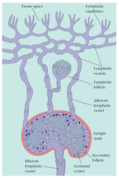
Figure 4. Lymphatic vessels. Small lymphatic capillaries opening into the tissue spaces pick up interstitial tissue fluid and carry it into progressively larger lymphatic vessels, which carry the fluid, now called lymph, into regional lymph nodes. As lymph leaves the nodes, it is carried through larger efferent lymphatic vessels, which eventually drain into the circulatory system at the thoracic duct or right lymph duct (see Figure 1).
The largest lymphatic vessel, the thoracic duct,empties into the left subclavian vein near the heart (see Figure 1). In this way, the lymphatic system captures fluid lost from the blood and returns it to the blood, thus ensuring steady-state levels of fluid within the circulatory system. The heart does not pump the lymph through the lymphatic system; instead the flow of lymph is achieved as the lymph vessels are squeezed by movements of the body’s muscles. A series of one-way valves along the lymphatic vessels ensures that lymph flows only in one direction.
When a foreign antigen gains entrance to the tissues, it is picked up by the lymphatic system (which drains all the tissues of the body) and is carried to various organized lymphoid tissues such as lymph nodes, which trap the foreign antigen. As lymph passes from the tissues to lymphatic vessels, it becomes progressively enriched in lymphocytes. Thus, the lymphatic system also serves as a means of transporting lymphocytes and antigen from the connective tissues to organized lymphoid tissues where the lymphocytes may interact with the trapped antigen and undergo activation.
Дата добавления: 2016-07-18; просмотров: 4149;
Поиск по сайту
Узнать еще
- Acquired immune deficiency syndrome (AIDS)
- Antibody-Antigen Interactions and the Function of the Fab
- Antibody-Antigen Interactions and the Function of the Fab
- Find the non-finite forms of the verb in the text and define their functions.
- Functions of the skeleton
- Glands: hormones and functions
- Intonation: approaches, definitions, functions
- Problems with the Immune System

Публикации по технике и механике

Публикации по биологии

Публикации по информатике

Публикации по строительству

Публикации по физике

Публикации по химии

Публикации по электронике

Публикации по искусству

Публикации по географии

Публикации по медицине
