Structure of bacteria and yeasts
Purpose of work:
To familiarize with the structure of bacteria, mushrooms and yeasts.
Materials and equipment:
Collection of cultures should to recultivate each 2-3 months on fresh nutrient media: bacteria and actinomycetes - on meat peptone agar, barmy and mold mushrooms on a wort-agar. 2 - 3 days before classes cultures recultivate in test tubes (bacteria and actinomycetes) or Petri dishes (mushrooms) from calculation after a bottom of a test tube of each culture and one-two Petri dishes on group of students of 10-15 persons. Microscopes and all accessories are necessary for classes; slide and cover glasses; bacteriological loops (needle); microscopic needles; a fresh 5 %-s' solution of fuchsine
Cells from animals, plants, and fungi are eukaryotes (Greek for "true nucleus"), whereas bacteria and the blue-green algae belong to the prokaryotes (Greek for "primitive nucleus"). In addition to lacking a nucleus and other organelles, prokaryotes use a smaller ribosome, the 70S ribosome, and in most bacteria, a mesh like peptidoglycan cell wall surrounds the membranes to protect it against the environment. Bacteria can survive and, in some cases, grow in hostile environments in which the osmotic pressure outside the cell is so low that most eukaryotic cells would lyse, at temperature extremes (both hot and cold), with dryness, and with very dilute and diverse energy sources. Bacteria have evolved the structures and functions to adapt to these conditions. Several of these distinctions provide the basis for antimicrobial action.
Bacteria are a large domain of prokaryotic microorganisms. Typically a few micrometres in length, bacteria have a wide range of shapes, ranging from spheres to rods and spirals. Bacteria are present in most habitats on Earth, growing in soil, acidic hot springs, radioactive waste, water, and deep in the Earth's crust, as well as in organic matter and the live bodies of plants and animals, providing outstanding examples of mutualism in the digestive tracts of humans, termites and cockroaches. There are typically 40 million bacterial cells in a gram of soil and a million bacterial cells in a millilitre of fresh water; in all, there are approximately five nonillion (5×1030) bacteria on Earth, forming a biomass that exceeds that of all plants and animals.
Bacteria can be distinguished from one another by their morphology (size, shape, and staining characteristics) and metabolic, antigenic, and genetic characteristics. Although bacteria are difficult to differentiate by size, they do have different shapes. A spherical bacterium, such as Staphylococcus, is a coccus; a rod-shaped bacterium, such as Escherichia coli, is a bacillus; and the snakelike treponeme is a spirillum. In addition, Nocardia and Actinomyces species have branched filamentous appearances similar to those of fungi. Some bacteria form aggregates such as the grapelike clusters of Staphylococcus aureus or the diplococcus (two cells together) observed in Streptococcus or Neisseria species.
The Gram stain is a powerful, easy test that allows clinicians to distinguish between the two major classes of bacteria and to initiate therapy. Bacteria that are heat-fixed or otherwise dried onto a slide are stained with crystal violet; this stain is precipitated with Gram iodine, and then the unbound and excess stain is removed by washing with the acetone-based decolorizer. A counterstain, safranin, is added to stain any decolorized cells red. This process takes less than 10 minutes.
For Gram positive bacteria, which turn purple, the stain gets trapped in a thick, cross-linked, meshlike structure, the peptidoglycan layer, which surrounds the cell. Gram negative bacteria have a thin peptidoglycan layer that does not retain crystal violet stain, so the cells must be counterstained with safranin and turned red. A mnemonic device that may help is "P-Purple-Positive." Gram stain is not a dependable test for bacteria that are starved (e.g., old or stationary phase cultures).
Bacteria that cannot be classified by Grain stain include mycobacteria, which have a waxy outer shell and are distinguished with the acid-fast stain, and mycoplasmas, which have no peptidoglycan.
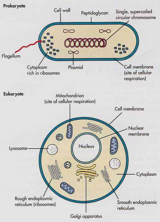
Figure 2. Major features of prokaryotes and eukaryotes.
Дата добавления: 2021-02-19; просмотров: 769;
Поиск по сайту
Узнать еще

Публикации по технике и механике

Публикации по биологии

Публикации по информатике

Публикации по строительству

Публикации по физике

Публикации по химии

Публикации по электронике

Публикации по искусству

Публикации по географии

Публикации по медицине
