Cutaneous-Associated Lymphoid Tissue
The skin is an important anatomic barrier to the external environment, and its large surface area makes this tissue important in nonspecific (innate) defenses. The epidermal (outer) layer of the skin is composed largely of specialized epithelial cells called keratinocytes. These cells secrete a number of cytokines that may function to induce a local inflammatory reaction. In addition, keratinocytes can be induced to express class II MHC molecules and may function as antigen-presenting cells. Scattered among the epithelial-cell matrix of the epidermis are Langerhans cells, a type of dendritic cell, which internalize antigen by phagocytosis or endocytosis. The Langerhans cells then migrate from the epidermis to regional lymph nodes, where they differentiate into interdigitating dendritic cells. These cells express high levels of class II MHC molecules and function as potent activators of naive TH cells.
The epidermis also contains so-called intraepidermal lymphocytes. These are similar to the intraepithelial lymphocytesof MALT in that most of them are CD8+ T cells, many ofwhich express γδ T-cell receptors, which have limited diversityfor antigen. These intraepidermal T cells are well situatedto encounter antigens that enter through the skin and someimmunologists believe that they may play a role in combatingantigens that enter through the skin. The underlying dermallayer of the skin contains scattered CD4+ and CD8+ Tcells and macrophages. Most of these dermal T cells were eitherpreviously activated cells or are memory cells.
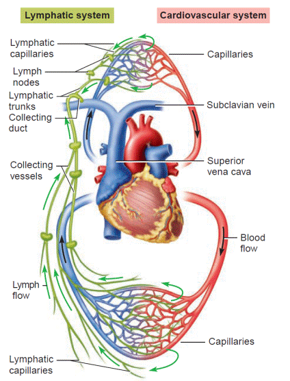
Figure. Comparison of the circulation patterns of the regular circulatory and lymphatic systems.The movement of lymphatic flow is in one direction (green) from lymph capillaries to collecting vessels and ducts to large lymphatic trunks to subclavian veins to the heart. The flow of blood on the other hand, is cyclic, with blood continuously fl owing through arteries to capillaries to veins to the heart and back around. With this combined system the lymphatics can collect excess tissue fluid and return it to the bloodstream. The two systems can also participate together in surveillance of the tissues for foreign invaders. Note: The lymphatic flow (green) is shown on only one side to keep the comparison uncluttered.
Дата добавления: 2016-07-18; просмотров: 2231;
Поиск по сайту
Узнать еще

Публикации по технике и механике

Публикации по биологии

Публикации по информатике

Публикации по строительству

Публикации по физике

Публикации по химии

Публикации по электронике

Публикации по искусству

Публикации по истории
